Imaging plays a crucial role in diagnosis management and follow-up for these patients and imaging technology over the last 100 years has advanced as the disease prevalence has. Such programmes have a number of components and their implementat.
What Is Fluoroscopy And How To Prepare Envision Radiology
An X-ray procedure allowing the visualization of internal organs in motion The meaning of the root blepharo is.

. A scan using an X-ray beam rotating around the patient Correct Answer. An instrument used chiefly in industry and in medical diagnosis for observing the internal structure of opaque objects as the living body by means of the shadow cast by the object. Technique using magnetism radio waves and a computer to produce images b.
Chapter 2 Anatomy and Medical Terminologypdf. Use of high-frequency sound waves to image anatomic structures. There are a few situations where a doctor might order a fluoroscopy test in Gainesville TX.
Radiographic positioning terminology is used routinely to describe the position of the patient for taking various radiographsStandard nomenclature is employed with respect to the anatomic position. The radiology term fluoroscopy is described as. This technique is used to image arteries in the brain heart kidneys gastrointestinal tract aorta neck carotids chest limbs and pulmonary circuit.
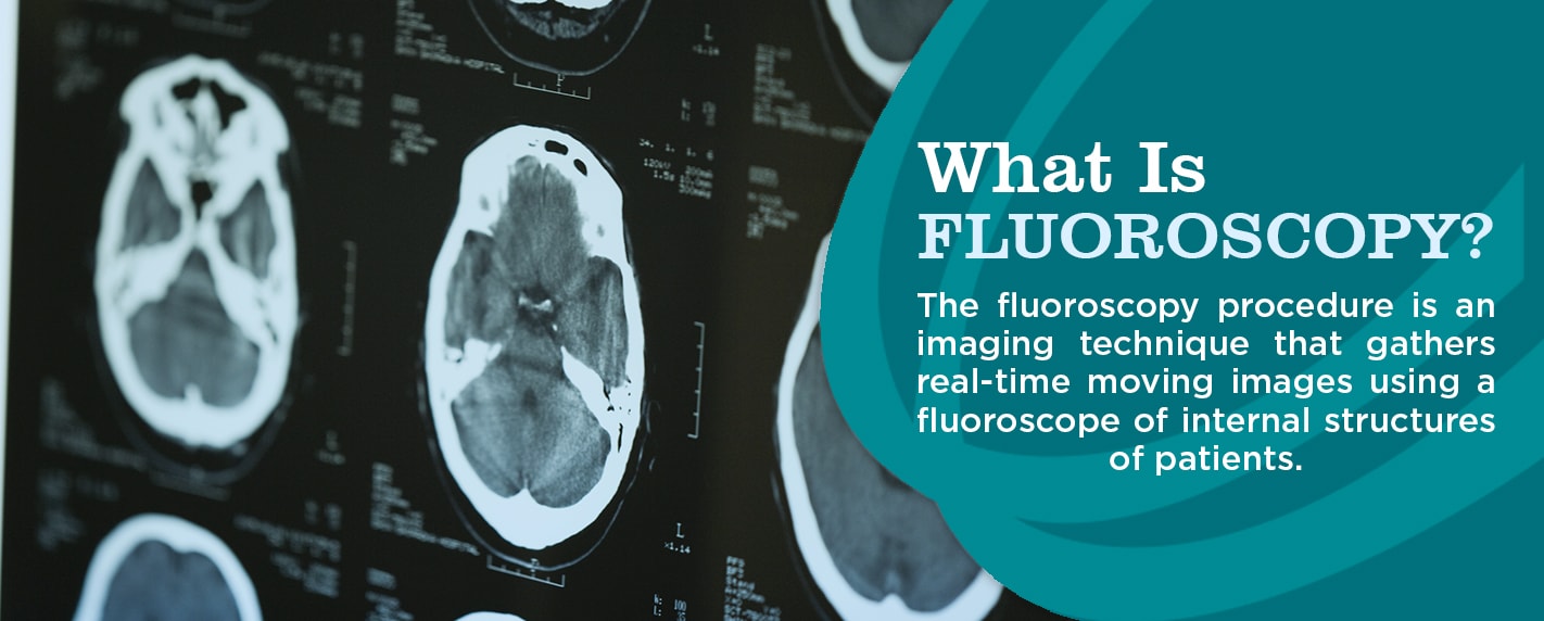
This term refers to structures or lesions becoming brighter on imaging after an intravenous contrast medium has been injected and can be used when contrast has been injected for a CT MRI or ultrasound. Programmes to manage patient dose in radiology are becoming a higher priority as the number of imaging examinations and the proportion of higher dose computed tomography CT and complex interventional procedures all continue to rise. An X-ray procedure allowing the visualization of internal organs in motion.
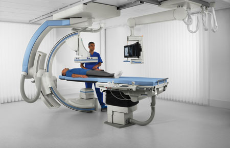
It is based on an x-ray image intensifier coupled to a stillvideo camera. The imaging is helpful to the doctor when inserting catheters wires and other small instruments and tools into your body. It is a unique and essential tool within diagnostic radiology and must remain an integral part of practice and residency training.
Anterior is towards the front of the body Latin. This preview shows page 10 out of 10 pages. This typically allows for smaller incisions cuts.
Terminology Basic terms of relations. An X-ray procedure allowing the visualization of internal organs in motion c. Today fluoroscopy is used foAcquisition parameters referencer diagnosis and guidance of clinical procedures.
A scan using an X-ray beam rotating around the patient d. Posterior is towards the back of the body Latin. A radiographic technique in which a radiopaque shows up on X-ray contrast material is injected into a blood vessel for the purpose of identifying its anatomy on an X-ray.
During a fluoroscopy procedure. Fluoroscopy Fluoroscopy utilizes a continuous beam of x-ray radiation to generate a moving picture image which is viewed on a computer monitor. Interventional radiologists are doctors that use imaging such as CT ultrasound MRI and fluoroscopy to help guide procedures.
Fluoroscopy can assist in evaluating issues with moving joints gastrointestinal issues lung. An X-ray procedure allowing the visualization of internal organs in motion c. Technique using magnetism radio waves and a computer to produce images b.
The prevalence of urinary stones in the United States has been described as 1 in 11 persons reporting a history of stones. Using X-ray technology fluoroscopy takes and displays several images of the inside of the body per second. Real time imaging viewed on a display monitor in the clinical room.
Matrix is smaller 512 x 512 pixels and 8 bits of grey scale only needed as temporal not spatial resolution is prioritised. The radiology term fluoroscopy is described as. Fluoroscopy Fluoroscopy is often described as an x-ray movie Its a continuous image that shows movement of a body part or motion of materials through the body.
Can acquire continuous cine or pulsed fluorographic images. The radiology term fluoroscopy is described as. Fluoroscopy remains a valuable modality even in the age of high-resolution cross-sectional imaging modalities such as computed tomography CT magnetic resonance imaging MRI and ultrasound.
Describe the functions of the main components of an X-ray tube including the cathode the anode the collimator and the filters. Fluoroscopy is a type of medical imaging that shows a continuous X-ray image on a monitor much like an X-ray movie. Fluoroscopy is an imaging system used by doctors to obtain a real time moving picture of the inside of the body.
This test can be thought of as a continuous X-ray that generates a movie as the doctor carries out the procedure. The radiology term fluoroscopy is described as. Fluoroscopy-guided catheter angiography is an interventional procedure that uses percutaneous access of arteries with needles and catheters to inject contrast for vessel opacification1 This procedure may be diagnostic or therapeutic.
Shortly after the discovery of X-rays in the late nineteenth century fluoroscopy was developed to enable visualization of moving anatomy. Applications of fluoroscopy are found throughout medicine including radiology cardiology. Fluoroscopy is a specific type of imaging scan that a doctor might order.
Describe the roles of an X-ray generator and its desired characteristics for medical imaging. In recent years flat panel detectors which are similar to the digital radiography used in projection radiography have been replacing the image intensifiers. Fluoroscopy is an imaging modality that allows real-time x-ray viewing of a patient with high temporal resolution.
What Is Fluoroscopy And How To Prepare Envision Radiology
Fluoroscopy Fda

Fluoroscopy Undergraduate Diagnostic Imaging Fundamentals


0 Comments